
Conditions · Spine surgery · Lainate
Biportal spinal endoscopy: spine surgery through two small incisions
The minimally invasive biportal endoscopic technique makes it possible to treat problems such as disc herniation and hypertrophy of the ligamentum flavum through two small incisions, with the aim of reducing muscle damage and supporting a quick recovery.
What it is
In biportal endoscopy (UBE, unilateral biportal endoscopy) the surgeon works through two incisions of about one centimetre: the endoscope, a small high-definition camera, goes through one, and the instruments through the other. The surgical field is continuously irrigated with saline solution, which keeps the view clear. The muscles are gently spread apart rather than cut.
What it is used for
- Lumbar disc herniation that does not respond to conservative treatment.
- Lumbar stenosis, particularly when a thickened ligamentum flavum compresses the nerves: removing it frees the canal.
- Foraminal stenosis, where the nerve roots exit the spine.
How it is performed
The operation is performed under general anaesthesia, with the patient lying face down. Under X-ray guidance, the two small incisions are made, the disc fragment or the tissue compressing the nerve is removed, and the surgeon checks that the nerve root is free. Patients are often out of bed just a few hours later; discharge and recovery times depend on each case and are discussed during the consultation.
What the studies say
In a multicentre randomised trial on lumbar disc herniation, biportal endoscopic discectomy proved non-inferior to microdiscectomy at 12 months, with slightly less wound pain in the first 48 hours and less muscle damage [1]. In lumbar stenosis, a meta-analysis found no significant differences in efficacy and safety compared with microsurgery, with a shorter average hospital stay [2]. The authors stress that more studies are needed: the technique should be chosen according to the individual case and the surgeon's experience.
Endoscopy or microsurgery?
Both are minimally invasive techniques with comparable results. The choice depends on the location and type of herniation or stenosis, the patient's anatomy and any previous surgery. Not every case is suitable for endoscopy: if there is abnormal movement between the vertebrae, aspinal fusion (arthrodesis).
After surgery
Proper rehabilitation is essential to recover function and reduce the risk of recurrence: post-operative physiotherapy strengthens the muscles and supports a gradual return to activities. With telemedicine, follow-up checks can also be done remotely.
Videos by Dr. Lavanga
Biportal endoscopy: the ultra-minimally invasive technique for disc herniation
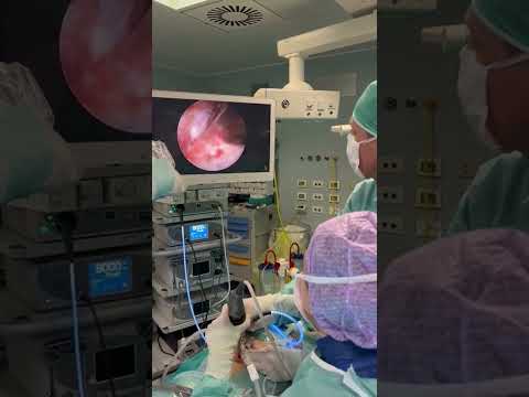 Watch on YouTube ↗Surgical images4:15
Disc herniation treated with biportal endoscopy: the surgical technique
Videos load from YouTube (youtube-nocookie.com) only when you press ▶.
Related articles
Scientific references (PubMed)
- Park SM et al. Biportal endoscopic versus microscopic discectomy for lumbar herniated disc: a randomized controlled trial. Spine J. 2023. PMID 36155241
- Pranata R et al. Biportal Endoscopic Spinal Surgery versus Microscopic Decompression for Lumbar Spinal Stenosis: A Systematic Review and Meta-Analysis. World Neurosurg. 2020. PMID 32147545
Information only: this does not replace a medical consultation, diagnosis or personalised treatment.