
Conditions · Spine surgery · Lainate
Herniated disc: symptoms, treatment and when to operate
A herniated disc occurs when part of an intervertebral disc moves out of place and irritates or compresses a nerve root. This causes pain running down the leg (sciatica) or down the arm, tingling and sometimes loss of strength. It is most common between the ages of 30 and 50 [6] and, in the vast majority of cases, it improves without surgery.
What the intervertebral disc is
Between one vertebra and the next there is a disc that acts as a shock absorber and allows the spine to bend and rotate. It is made up of an outer annulus fibrosus , made of layers of criss-crossing fibres, and an inner nucleus pulposus , which is gel-like and rich in water. Behind the disc runs the spinal canal, from which the nerve roots leading to the legs emerge (Figure 1).
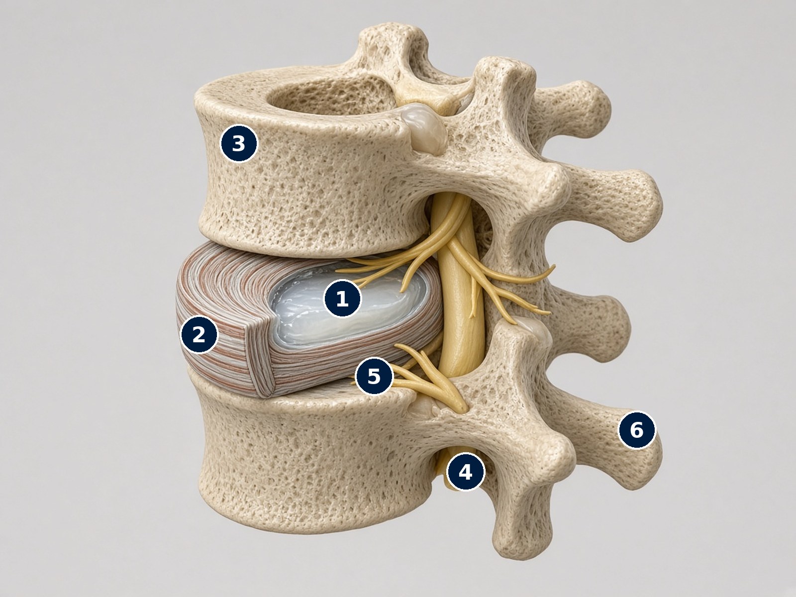
The healthy disc and the nerve roots
Two lumbar vertebrae with the disc between them: a wedge has been removed to show the inside.
Side view, slightly from behind: on the left is the front of the spine (abdomen), on the right the back.
What each number shows
- 1Nucleus pulposus. The central, water-rich gel. Under load it changes shape and spreads pressure evenly, like a hydraulic cushion.
- 2Annulus fibrosus. Concentric layers of criss-crossing fibres that contain the nucleus. With age and repeated loading, small tears can appear: this is where a herniation starts.
- 3Vertebral body. The front and strongest part of the vertebra, which bears the weight. The disc is firmly attached to the endplates above and below and cannot “slip out”.
- 4Dural sac. The sheath containing the cerebrospinal fluid and, in the lumbar region, the nerve roots of the cauda equina (the spinal cord itself ends higher up).
- 5Nerve roots. They branch off the dural sac and exit sideways and backwards through the intervertebral foramina, beneath the pedicle of each vertebra. They carry sensation and strength to the leg.
- 6Posterior elements. Spinous processes, transverse processes and facet joints: they protect the spinal canal, guide movement and provide attachment for muscles.
Simplified illustration: the roots are drawn slightly fanned out to make them visible.
How a herniation forms
With age the disc loses water and the annulus can tear. Under load, part of the nucleus pushes outwards. International nomenclature distinguishes three main forms [7]:
- Protrusion: the disc bulges beyond its margin with a broad base; the annulus still contains it.
- Extrusion (expelled herniation): the material passes through the tear and its outer part is wider than the “neck” it came out of.
- Sequestration (migrated herniation): a fragment breaks away completely from the disc and can move up or down within the canal.
Pain does not depend only on mechanical compression: the nucleus material also causesinflammation of the nerve root. This is why a small herniation can be very painful and a large one, at times, almost painless.
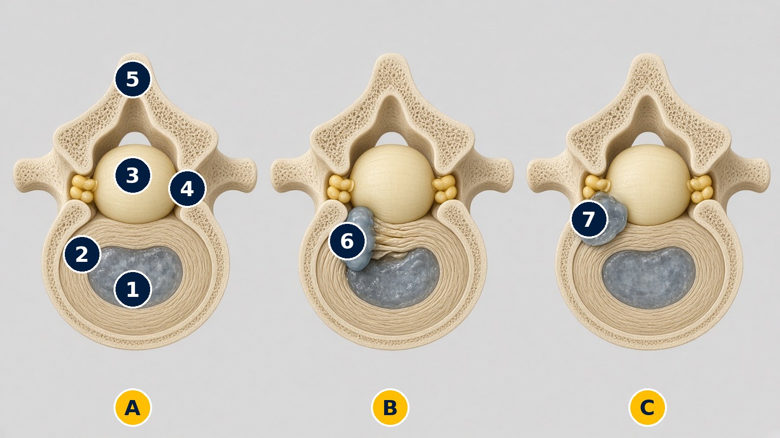
How a herniation forms: from the intact disc to the migrated fragment
The same lumbar vertebra seen from above in three different situations.
Horizontal section seen from above: at the bottom is the front (the disc), at the top the back (the bony arch and the spinal canal).
What each number shows
- AIntact disc. The nucleus is contained by the annulus and the canal is clear. In a protrusion, the first stage, the back edge of the disc bulges only slightly with a broad base, but the annulus remains intact [7].
- BExpelled herniation (extrusion). The nucleus material passes through a tear in the annulus, sideways and backwards (posterolateral), and enters the canal, pressing on the dural sac and on the nerve root on that side [7].
- CMigrated herniation (sequestration). A fragment breaks away completely from the disc and lies free in the canal, next to the nerve root. Paradoxically, these are precisely the herniations that most often reabsorb [4].
- 1Nucleus pulposus. The central gel of the disc.
- 2Annulus fibrosus. The rings of fibres surrounding the nucleus.
- 3Dural sac. Contains the cerebrospinal fluid and the roots of the cauda equina.
- 4Nerve roots. On either side of the sac, heading towards the exit foramina.
- 5Posterior arch. Laminae and spinous process: they close the canal at the back.
- 6Expelled material. Nucleus that has come out through the tear, still connected to the disc.
- 7Free fragment. A piece of disc that has broken off, lying in the canal next to the nerve root.
Panel A shows the intact disc for comparison; the protrusion is described in the text because it would be barely visible in the drawing.
Symptoms: they depend on which nerve root is involved
Lumbar disc herniation mainly affects the L4-L5 and L5-S1 [6]levels. The typical symptom is sciatica: pain that runs from the back or buttock down the leg, often stronger than the back pain itself, and gets worse when sitting, coughing or sneezing.
| Level and root | Where it is felt | What may become weak |
|---|---|---|
| L4-L5 → L5 root | Outer side of the leg, top of the foot, big toe | Lifting the foot and big toe (walking on your heels) |
| L5-S1 → S1 root | Back of the thigh and calf, sole and outer edge of the foot | Standing on tiptoe; reduced Achilles reflex |
| Lateral L4-L5 herniation → L4 root | Front of the thigh, inner side of the leg | Straightening the knee; reduced knee-jerk reflex |
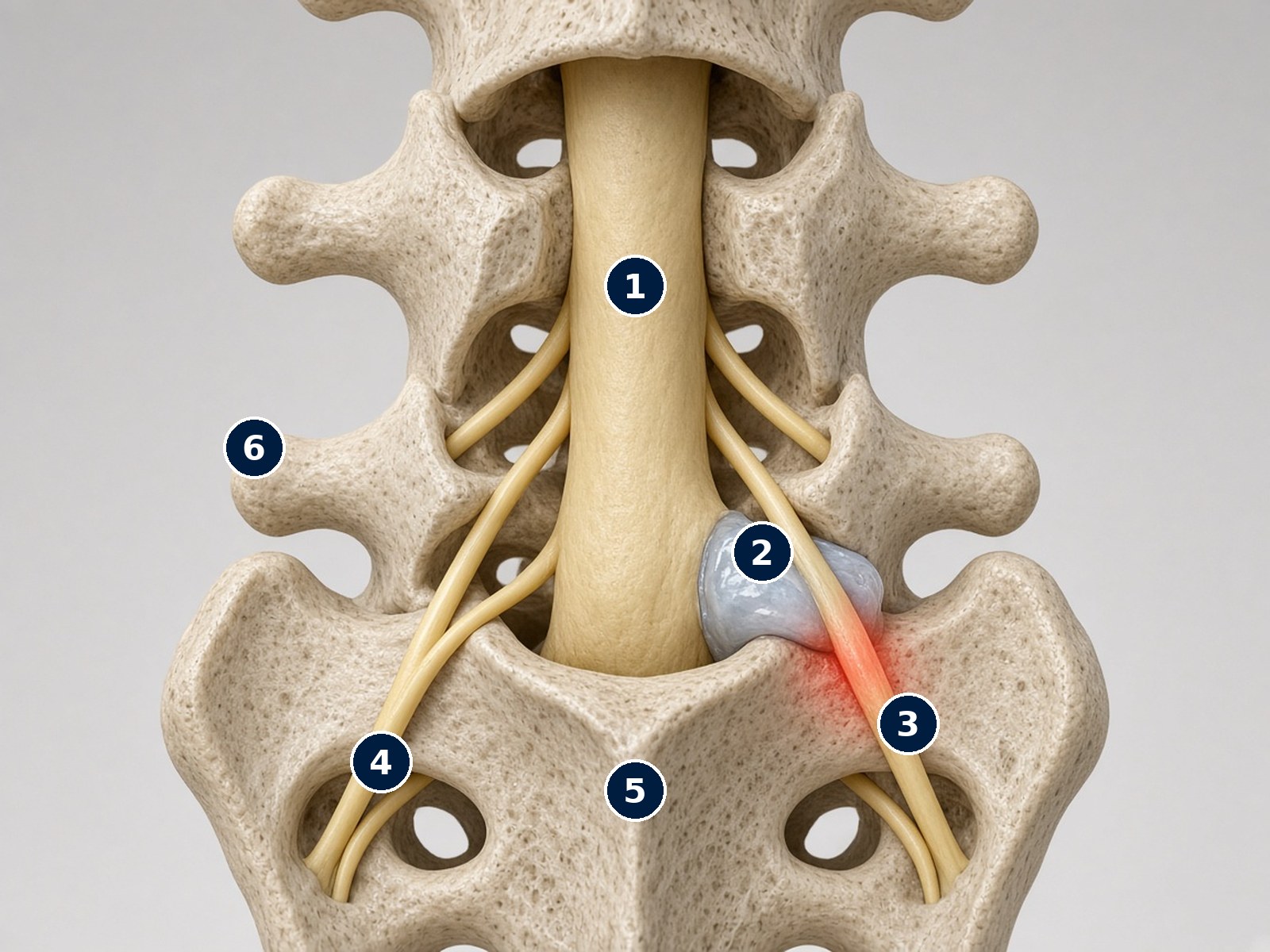
Which nerve is compressed: the example of an L5-S1 herniation
Lumbar spine seen from behind, with the posterior arch removed: a right-sided L5-S1 herniation compresses the S1 root.
Rear view: the laminae and spinous processes have been removed to show the inside of the canal. The patient's right corresponds to the right of the image.
What each number shows
- 1Dural sac. It runs down the middle of the canal; the nerve roots branch off its sides, level by level.
- 2Posterolateral L5-S1 herniation. Disc material between the fifth lumbar vertebra and the sacrum comes out sideways and backwards, next to the sac.
- 3Compressed S1 root. The root that has just branched off the sac and “passes over” the disc is compressed and inflamed (red halo). It causes pain in the back of the thigh and calf, the sole and outer edge of the foot, a reduced Achilles reflex and difficulty standing on tiptoe.
- 4Roots on the opposite side. They are free: this is why symptoms usually affect only one leg.
- 5Sacrum. The base of the spine; the sacral roots exit through its foramina.
- 6L5 transverse process. Lateral bony landmark of the last lumbar vertebra.
In the same way, a posterolateral L4-L5 herniation usually compresses the L5 root, while a lateral (foraminal) herniation compresses the root exiting at that level, i.e. L4. This is why the clinical examination, which identifies the root involved, must always match the MRI.
Cervical disc herniation. In the neck, a herniation causes pain radiating into the arm (cervicobrachialgia), with tingling in the fingers. The root most often involved is C7 (middle finger, triceps), followed by C6 (thumb, biceps) [11]. In a population-based study, years later 90% of people had no symptoms or only mild complaints [11]. Clumsiness when using the hands or an unsteady gait may indicate that the spinal cord is under strain (myelopathy) and require a prompt consultation. Read more: cervicobrachialgia.
When to go straight to the Emergency Department
Please do not wait for your scheduled appointment if you notice:
- difficulty holding or passing urine or stools;
- “saddle” numbness (genital area, perineum, inner thighs);
- loss of strength that is getting rapidly worse in one or both legs.
These may indicate cauda equina syndrome, a severe compression of all the lumbar nerve roots [5]. It is a surgical emergency: a meta-analysis showed better outcomes for sensation, strength, bladder and bowel when decompression is performed within 48 hours of onset [10]. Fever, a history of cancer or a significant injury associated with back pain also require prompt assessment [5].
How it is diagnosed
Diagnosis starts with the clinical examination: the description of symptoms, the straight-leg raise test (Lasègue), reflexes, strength and sensation show which nerve root is involved. Magnetic resonance imaging (MRI) is the reference test when symptoms are consistent with radiculopathy [3]: it confirms the herniation and shows its size and position (Figure 4). CT and electromyography are used in selected cases. Without warning signs, an MRI is not always needed straight away: it is requested if symptoms do not improve or if an injection or surgery is being considered.
A word of caution about reports. In a study of 98 people without back pain, 52% had a disc bulge, 27% a protrusion and 1% an extrusion [8]. A review of 3,110 pain-free people found protrusions in 29% of people in their twenties and 43% of those in their eighties [9]. A “herniation” on a report therefore matters only if it matches the symptoms and the clinical examination.
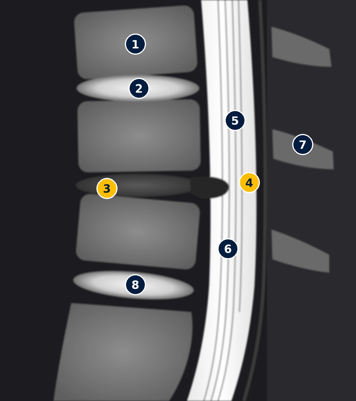
What a herniation looks like on MRI
Diagram of a lumbar MRI in side (sagittal) view, T2 sequence: water appears white.
Teaching diagram, not a patient's MRI. On the left is the front, on the right the back; from top to bottom L3, L4, L5 and the sacrum.
What each number shows
- 1Vertebral body. The vertebrae appear grey, with a uniform signal.
- 2Healthy disc (L3-L4). Bright on T2 because it is rich in water.
- 3Degenerated disc (L4-L5). Darker and flatter (the so-called “black disc”): it has lost water. This is a very common finding, even in people without pain [9].
- 4Herniation. A small dark bulge behind the disc that enters the canal and “interrupts” the white column of cerebrospinal fluid.
- 5Cerebrospinal fluid. The fluid surrounding the nerve roots: on T2 it is bright white and acts as a natural contrast.
- 6Cauda equina roots. The thin lines running down through the fluid.
- 7Spinous processes. The posterior bony elements, beneath the skin of the back.
- 8Healthy disc (L5-S1). Bright again, with preserved height.
An MRI must always be read together with the symptoms: protrusions and dark discs are often found in people with no pain at all.
Does a herniation reabsorb on its own?
Often, yes. The immune system recognises the material that has come out of the disc as foreign and gradually “clears it away”. A systematic review calculated spontaneous regression in 96% of migrated herniations, in 70% of extruded herniations, in 41% of protrusions and in 13% of simple bulges; complete resolution occurred in 43% of sequestrations and 15% of extrusions [4]. The largest herniations, those that have “come out”, are therefore not necessarily the worst.
Can it heal without surgery?
Yes, in most cases. Conservative treatment includes:
- Information and movement: staying active within the limits of pain; prolonged bed rest does not help and weakens the muscles.
- Medication: painkillers and anti-inflammatories, as prescribed by the doctor and for as long as needed.
- Physiotherapy: personalised exercises to control pain, recover movement and strengthen the trunk.
- Injections: in selected cases, cortisone injected near the nerve root (epidural or periradicular, under imaging guidance) can provide short-term relief [3].
The treatment pathway for a herniated disc
Conservative treatment
First weeks
- Information and reassurance: a herniation often shrinks on its own [4]
- Stay active, avoid prolonged bed rest
- Medication as prescribed by the doctor
- Physiotherapy and guided exercise
Move to the next step if pain remains severe and disabling despite well-conducted treatment.
Injection
Selected cases
- Cortisone near the inflamed nerve root (epidural or periradicular), under imaging guidance
- Goal: short-term relief to get through the acute phase [3]
Surgery is considered if the benefit is insufficient or short-lived.
At every stage, the MRI must match the clinical examination. We do not operate on an image, we treat a patient.
When surgery is considered
Surgery is recommended in case of significant or worsening loss of strength, cauda equina syndrome or disabling leg pain that does not improve after about 6–12 weeks of well-conducted treatment, with an MRI that matches the symptoms.
What the most important studies say:
- In the Dutch study published in the New England Journal of Medicine, patients operated on early had faster relief from leg pain; at one year, however, the likelihood of feeling recovered was 95% in both groups. In the conservative group, 39% eventually had surgery over time [2].
- In the American SPORT study, both groups improved substantially [1]; at 8 years, those who had surgery showed greater improvement, with stable results over time in both groups [12].
The decision is therefore not “dictated” by the image: it is made together with the patient, based on the intensity of the pain, their work, their expectations and how quickly they wish to return to normal life.
How microdiscectomy works
The most common operation is microdiscectomy: through a small incision and using a microscope, a small window is opened in the bone, the nerve root is gently moved aside and only the fragment compressing it is removed. The rest of the disc stays in place and no screws are inserted (Figure 5). In selected cases Dr. Lavanga usesbiportal spinal endoscopy, through two small incisions.
Endoscopic techniques have been compared with traditional microdiscectomy: in a randomised trial of 613 patients, transforaminal endoscopy proved non-inferior, with less blood loss, a shorter hospital stay and earlier mobilisation [13].
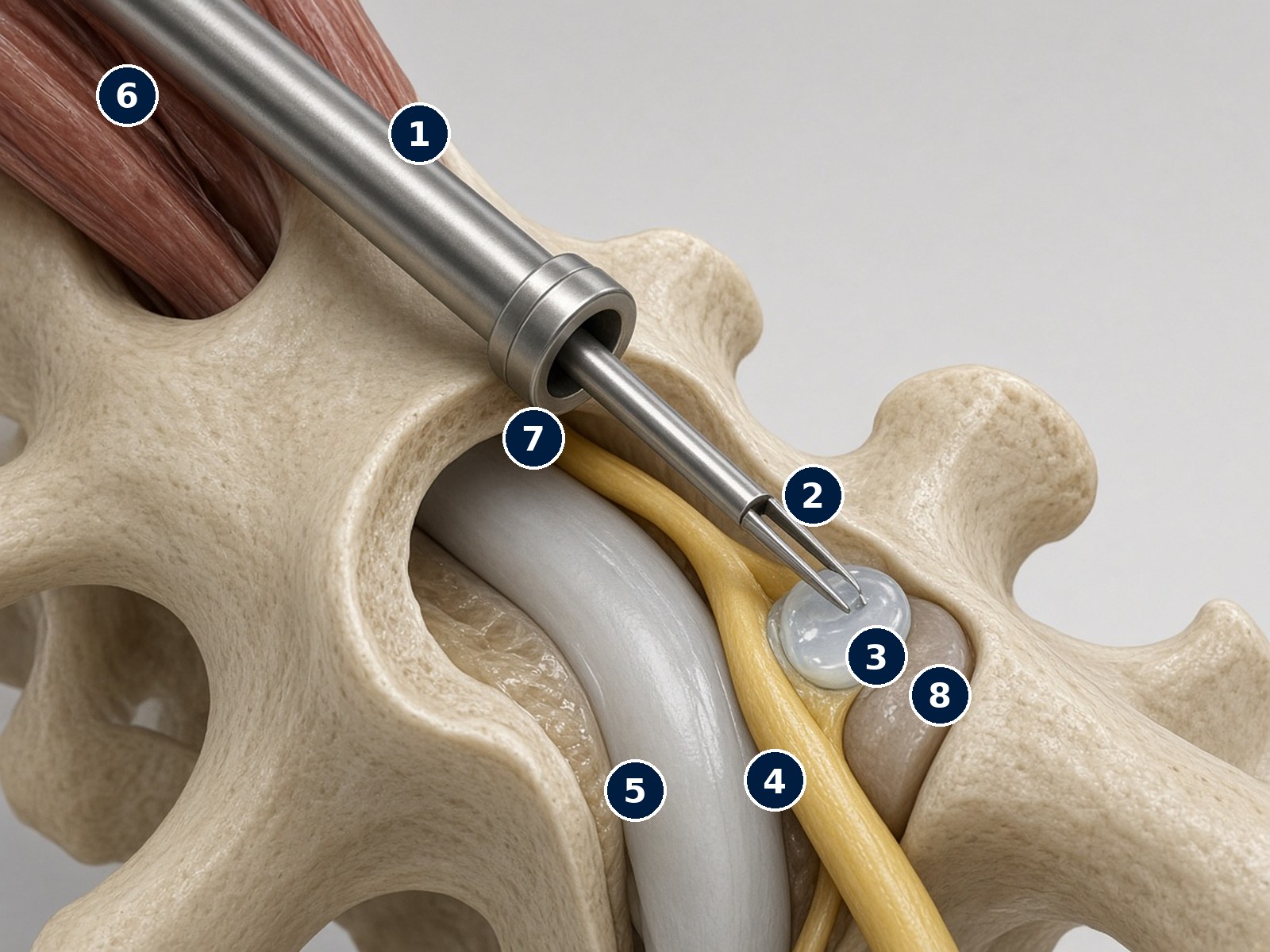
Microdiscectomy: only the fragment is removed
The concept of minimally invasive microdiscectomy: through a small opening, the fragment compressing the nerve root is removed.
Oblique rear view of the lumbar spine during surgery.
What each number shows
- 1Minimally invasive access. A small retractor through an incision of a few centimetres; the surgeon works with the microscope or, in selected cases, with the endoscope.
- 2Micro-forceps. Grasps and removes the herniated fragment.
- 3Herniated fragment. The part of the disc that has moved out of place: it is the only part removed.
- 4Nerve root. Once the pressure is relieved, leg pain usually eases quickly.
- 5Dural sac. It is protected and gently moved aside.
- 6Paraspinal muscles. They are spread apart, not cut.
- 7Small bony window. Only the space needed is opened at the edge of the lamina (laminotomy).
- 8Disc. The rest of the disc stays in place: no screws or implants are used; this is not a spinal fusion.
Illustration of the principle: the exact technique (microscope, tubular retractor or biportal endoscopy) is chosen case by case.
Recovery after surgery
- Same day or next day: you get up and walk; leg pain usually eases within the first few days, while tingling and strength may take weeks or months to recover.
- First weeks: daily walks of increasing length, avoiding lifting heavy objects and sitting for long periods.
- Work and sport: desk jobs are usually resumed earlier than physically demanding ones; timing is agreed at the follow-up consultation.
- Physiotherapy: a trunk-strengthening programme with the Ortho-Next physiotherapists supports a gradual return to activities.
Can the herniation come back?
In a minority of cases, yes. In the Dutch randomised trial on endoscopy, a repeat operation was needed within one year in 5–6% of patients, with similar results for the two techniques [13]. Weight control, trunk strengthening and a gradual return to loading help reduce the risk. For repeated recurrences, or when a worn disc is combined with a failing segment, alumbar spinal fusionmay be considered, for example with the TLIF technique.
Prevention
Regular physical activity, frequent breaks from sitting, correct lifting technique (straight back, load close to the body, power from the legs), maintaining a healthy weight and stopping smoking all support disc health and reduce the risk of relapse.
Videos by Dr. Lavanga
Lumbar disc herniation: what it is and what it causes
Herniated disc: the symptoms not to ignore and when surgery is needed
Herniated disc: spinal orthopaedics
Low back pain with sciatica: understanding the pain and how to treat it
Radio Dossier24: herniated disc, symptoms, diagnosis and modern treatment
Videos load from YouTube (youtube-nocookie.com) only when you press ▶.
Related articles
Scientific references (PubMed)
- Weinstein JN et al. Surgical vs nonoperative treatment for lumbar disk herniation: the Spine Patient Outcomes Research Trial (SPORT): a randomized trial. JAMA. 2006. PMID 17119140
- Peul WC et al. Surgery versus prolonged conservative treatment for sciatica. N Engl J Med. 2007. PMID 17538084
- Kreiner DS et al. An evidence-based clinical guideline for the diagnosis and treatment of lumbar disc herniation with radiculopathy. Spine J. 2014. PMID 24239490
- Chiu CC et al. The probability of spontaneous regression of lumbar herniated disc: a systematic review. Clin Rehabil. 2015. PMID 25009200
- Finucane LM et al. International Framework for Red Flags for Potential Serious Spinal Pathologies. J Orthop Sports Phys Ther. 2020. PMID 32438853
- Jordan J, Konstantinou K, O'Dowd J. Herniated lumbar disc. BMJ Clin Evid. 2009. PMID 19445754
- Fardon DF et al. Lumbar disc nomenclature: version 2.0. Recommendations of the combined task forces of NASS, ASSR and ASNR. Spine J. 2014. PMID 24768732
- Jensen MC et al. Magnetic resonance imaging of the lumbar spine in people without back pain. N Engl J Med. 1994. PMID 8208267
- Brinjikji W et al. Systematic literature review of imaging features of spinal degeneration in asymptomatic populations. AJNR Am J Neuroradiol. 2015. PMID 25430861
- Ahn UM et al. Cauda equina syndrome secondary to lumbar disc herniation: a meta-analysis of surgical outcomes. Spine. 2000. PMID 10851100
- Radhakrishnan K et al. Epidemiology of cervical radiculopathy. A population-based study from Rochester, Minnesota, 1976 through 1990. Brain. 1994. PMID 8186959
- Lurie JD et al. Surgical versus nonoperative treatment for lumbar disc herniation: eight-year results for the Spine Patient Outcomes Research Trial. Spine. 2014. PMID 24153171
- Gadjradj PS et al. Full endoscopic versus open discectomy for sciatica: randomised controlled non-inferiority trial. BMJ. 2022. PMID 35190388
Information only: this does not replace a medical consultation, diagnosis or personalised treatment. Illustrations for educational purposes. Last reviewed: October 2026.